43 the human heart and its labels
Heart - Wikipedia The human heart is situated in the mediastinum, at the level of thoracic vertebrae T5-T8.A double-membraned sac called the pericardium surrounds the heart and attaches to the mediastinum. The back surface of the heart lies near the vertebral column, and the front surface sits behind the sternum and rib cartilages. The upper part of the heart is the attachment point for several large blood ... Heart: illustrated anatomy - e-Anatomy - IMAIOS This interactive atlas of human heart anatomy is based on medical illustrations and cadaver photography. The user can show or hide the anatomical labels which provide a useful tool to create illustrations perfectly adapted for teaching. Anatomy of the heart: anatomical illustrations and structures, 3D model and photographs of dissection.
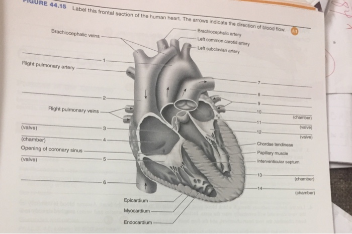
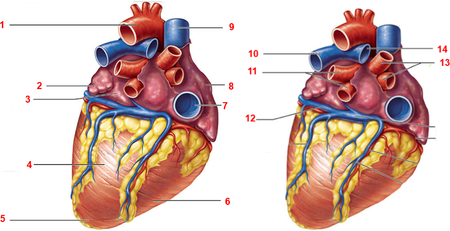
The 18 parts of the human heart, and their functions The 18 parts of the human heart and how they work 1. Myocardium 2. Endocardium 3. Pericardium 4. Right Auricle 5. Right ventricle 6. Tricuspid valve 7. Pulmonary valve 8. Left Auricle 9. Left ventricle 10. Mitral valve 11. Aortic valve 12. Tendon cords 13. Papillary muscles 14. Sinus node 15. Atrioventricular node 16. Atrioventricular fascicule 17.

The human heart and its labels
Heart: Anatomy and Function - Cleveland Clinic Heart. Your heart is the main organ of your cardiovascular system, a network of blood vessels that pumps blood throughout your body. It also works with other body systems to control your heart rate and blood pressure. Your family history, personal health history and lifestyle all affect how well your heart works. Appointments 800.659.7822. 147 Heart Anatomy With Labels Premium High Res Photos - Getty Images Browse 147 heart anatomy with labels stock photos and images available, or start a new search to explore more stock photos and images. of 3. NEXT. PDF Analyzing the Human Heart - Beyond the Classroom working. Its job is to pump blood to the lungs and to all of the body tissues. In this activity you will use a diagram of the heart to analyze the way in which the heart works. l. Using the following word list, label the various parts of the heart on the diagram. Right ventricle Left venfficle Upper vena cava Lower vena cava Aorta
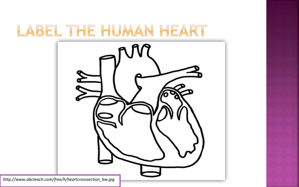
The human heart and its labels. Label the heart — Science Learning Hub In this interactive, you can label parts of the human heart. Drag and drop the text labels onto the boxes next to the diagram. Selecting or hovering over a box will highlight each area in the diagram. Aorta Vena cava Right ventricle Semilunar valve Left atrium Left ventricle Right atrium Pulmonary vein Pulmonary artery Download Exercise Tweet heart | Structure, Function, Diagram, Anatomy, & Facts heart, organ that serves as a pump to circulate the blood. It may be a straight tube, as in spiders and annelid worms, or a somewhat more elaborate structure with one or more receiving chambers (atria) and a main pumping chamber (ventricle), as in mollusks. In fishes the heart is a folded tube, with three or four enlarged areas that correspond to the chambers in the mammalian heart. In animals ... 13 parts of the human heart (and its functions) - LORECENTRAL Parts of the heart and its functions 1. Left atrium 2. Mitral Valve 3. Left Ventricle 4. Aortic sigmoid valve Right atrium 6. Tricuspid valve 7. Right ventricle 8. Pulmonary sigmoid valve 9. Atrial septal defect Interventricular partition 11. The sinus or sinoatrial node 12. Atrioventricular or Aschoff-Tawara nodule 13. Hiscules and Purkinje fibers Human Heart Diagram - Human Body Pictures - Science for Kids Find free pictures, photos, diagrams, images and information related to the human body right here at Science Kids. Photo name: Human Heart Diagram Picture category: Human Body Image size: 70 KB Dimensions: 600 x 600 Photo description: This is an excellent human heart diagram which uses different colors to show different parts and also labels a number of important heart component such as the ...
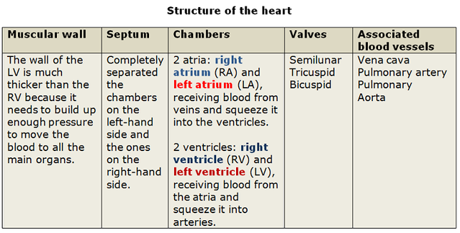
Parts Of The Human Heart | Science Trends The parts of the human heart can be broken down into four chambers, muscular walls, vessels, and a conductive system. The two upper chambers are called the atria, with lower parts called ventricles. These all work together to make up the vital function of your heart. Everybody knows that the human heart is the essential organ in our bodies. Anatomy of a Human Heart - U of M Health Located between the lungs in the middle of the chest, the heart pumps blood through the network of arteries and veins known as the cardiovascular system. It pushes blood to the body's organs, tissues and cells. Blood delivers oxygen and nutrients to every cell and removes the carbon dioxide and other waste products made by those cells. The Anatomy of the Heart, Its Structures, and Functions - ThoughtCo The heart is the organ that helps supply blood and oxygen to all parts of the body. It is divided by a partition (or septum) into two halves. The halves are, in turn, divided into four chambers. The heart is situated within the chest cavity and surrounded by a fluid-filled sac called the pericardium. This amazing muscle produces electrical ... parts of the heart label Label The Heart Diagram - Human Anatomy tartrerepub.blogspot.com. heart diagram worksheet blank label worksheets sparklebox human anatomy related. Lab 2 Pig Heart-Labeled | Cardiovascular, Estudos . heart anatomy lab pig physiology human labeled dissection sheep posterior pigs body salvo escolha pasta kctcs bluegrass district edu.
Human Heart (Anatomy): Diagram, Function, Chambers, Location in ... - WebMD The heart is a muscular organ about the size of a fist, located just behind and slightly left of the breastbone. The heart pumps blood through the network of arteries and veins called the... Diagram of Human Heart and Blood Circulation in It Exterior of the Human Heart A heart diagram labeled will provide plenty of information about the structure of your heart, including the wall of your heart. The wall of the heart has three different layers, such as the Myocardium, the Epicardium, and the Endocardium. Here's more about these three layers. Epicardium Diagram of the human heart royalty-free images - Shutterstock Find Diagram of the human heart stock images in HD and millions of other royalty-free stock photos, illustrations and vectors in the Shutterstock collection. Thousands of new, high-quality pictures added every day. How the Heart Works - The Heart | NHLBI, NIH - National Institutes of ... The heart is an organ about the size of your fist that pumps blood through your body. It is made up of multiple layers of tissue. Your heart is at the center of your circulatory system. This system is a network of blood vessels, such as arteries, veins, and capillaries, that carries blood to and from all areas of your body.
Anatomy of the Human Heart Quiz
Human heart: Anatomy, function & facts | Live Science The human heart has four chambers: two upper chambers (the atria) and two lower ones (the ventricles), according to the National Institutes of Health. The right atrium and right ventricle together...
Structure and function of the heart - Biology Notes for IGCSE 2014
13+ Heart Diagram Templates - Sample, Example, Format Download Human heart is a complicated figure and for students from science, they will often need the images of the heart for its illustration. The above collection of heart samples will make it easier for students to download, print and use it in their projects. The images with labels and detailed explanations can also be used in text books.
PPT - The Transport System PowerPoint Presentation, free download - ID:2209825
Human Heart - Anatomy, Functions and Facts about Heart - BYJUS The human heart is divided into four chambers, namely two ventricles and two atria. The ventricles are the chambers that pump blood and atrium are the chambers that receive the blood. Among which, the right atrium and ventricle make up the "right portion of the heart", and the left atrium and ventricle make up the "left portion of the heart." 5.
32 Label The Human Heart - Labels Design Ideas 2020
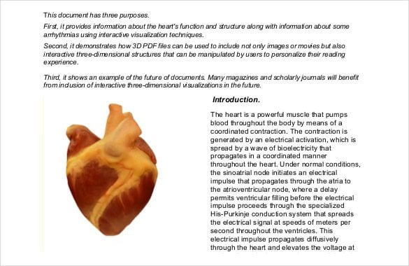
Labelling the heart — Science Learning Hub Labelling the heart — Science Learning Hub Labelling the heart Add to collection The heart is a muscular organ that pumps blood through the blood vessels of the circulatory system. Blood transports oxygen and nutrients to the body. It is also involved in the removal of metabolic wastes. Topics Concepts Citizen science Teacher PLD Glossary Sign in

Heart Diagram - 15+ Free Printable Word, Excel, EPS, PSD Template ... Teachers and students use the heart diagram, in biological science, to study the structure and functions of a human being's heart. ... Label The Parts Of The Heart. depts.washington.edu | Having the heart diagram for studies or for scientific purpose has been made easy through this template. It shows a heart picture with all its parts labeled ...
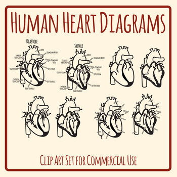
Human Heart - Label the Diagram Anatomy Clip Art Set Commercial Use
How the Heart Works: Diagram, Anatomy, Blood Flow - MedicineNet The heart is an amazing organ. It starts beating about 22 days after conception and continuously pumps oxygenated red blood cells and nutrient-rich blood and other compounds like platelets throughout your body to sustain the life of your organs.; Its pumping power also pushes blood through organs like the lungs to remove waste products like CO2.; This fist-sized powerhouse beats (expands and ...

Human digestive system diagram & function explained
Heart Diagram with Labels and Detailed Explanation - BYJUS Diagram of Heart. The human heart is the most crucial organ of the human body. It pumps blood from the heart to different parts of the body and back to the heart. The most common heart attack symptoms or warning signs are chest pain, breathlessness, nausea, sweating etc. The diagram of heart is beneficial for Class 10 and 12 and is frequently ...
13+ Heart Diagram Templates – Sample, Example, Format Download | Free & Premium Templates
How to Draw a Human Heart: 11 Steps (with Pictures) - wikiHow Label the parts of the heart if you'd to reference it for anatomy. If you're trying to identify parts of the heart for a class you're taking, it's good practice to draw the heart yourself and label each segment. You can refer to your textbook in order to label the: [9] Aorta Superior vena cava Inferior vena cava Right and left atria

Heart Anatomy Quizzes and Flashcards – sciencemusicvideos
File:Diagram of the human heart (no labels).svg File:Diagram of the human heart (no labels).svg. From Wikimedia Commons, the free media repository. File. File history. File usage on Commons. Metadata. Size of this PNG preview of this SVG file: 498 × 599 pixels. Other resolutions: 199 × 240 pixels | 399 × 480 pixels | 639 × 768 pixels | 851 × 1,024 pixels | 1,703 × 2,048 pixels | 533 × ...
Labeling the Heart (Part Two) Quiz - By dilatory
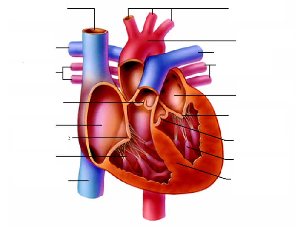
A Labeled Diagram of the Human Heart You Really Need to See The human heart, comprises four chambers: right atrium, left atrium, right ventricle and left ventricle. The two upper chambers are called the left and the right atria, and the two lower chambers are known as the left and the right ventricles. The two atria and ventricles are separated from each other by a muscle wall called 'septum'.

Label The Parts Of A Human Heart - Human Heart Diagram Images Stock Photos Vectors Shutterstock ...
A Diagram of the Heart and Its Functioning Explained in Detail The heart blood flow diagram (flowchart) given below will help you to understand the pathway of blood through the heart.Initial five points denotes impure or deoxygenated blood and the last five points denotes pure or oxygenated blood. 1.Different Parts of the Body ↓ 2.Major Veins ↓ 3.Right Atrium ↓ 4.Right Ventricle ↓ 5.Pulmonary Artery ↓ 6.Lungs

Quotes about Cardiovascular system (40 quotes)
Human Heart - Diagram and Anatomy of the Heart - Innerbody The heart contains 4 chambers: the right atrium, left atrium, right ventricle, and left ventricle. The atria are smaller than the ventricles and have thinner, less muscular walls than the ventricles. The atria act as receiving chambers for blood, so they are connected to the veins that carry blood to the heart.

PDF Analyzing the Human Heart - Beyond the Classroom working. Its job is to pump blood to the lungs and to all of the body tissues. In this activity you will use a diagram of the heart to analyze the way in which the heart works. l. Using the following word list, label the various parts of the heart on the diagram. Right ventricle Left venfficle Upper vena cava Lower vena cava Aorta
Heart Diagram Unlabeled - Cliparts.co
147 Heart Anatomy With Labels Premium High Res Photos - Getty Images Browse 147 heart anatomy with labels stock photos and images available, or start a new search to explore more stock photos and images. of 3. NEXT.

ARJUN'S CORNER......: KONSEP 10S
Heart: Anatomy and Function - Cleveland Clinic Heart. Your heart is the main organ of your cardiovascular system, a network of blood vessels that pumps blood throughout your body. It also works with other body systems to control your heart rate and blood pressure. Your family history, personal health history and lifestyle all affect how well your heart works. Appointments 800.659.7822.


i heart manila: manila ocean park - bahura fish pictures
Post a Comment for "43 the human heart and its labels"