45 microscope diagram with labels and definitions
Simple Microscope - Diagram (Parts labelled), Principle, Formula and Uses A simple microscope consists of Optical parts Mechanical parts Labeled Diagram of simple microscope parts Optical parts The optical parts of a simple microscope include Lens Mirror Eyepiece Lens A simple microscope uses biconvex lens to magnify the image of a specimen under focus. Simple Microscope - Parts, Functions, Diagram and Labelling Parts of the optical parts are as follows: Mirror - A simple microscope has a plano-convex mirror and its primary function is to focus the surrounding light on the object being examined. Lens - The biconvex lens is placed above the stage and its function is to magnify the size of the object being examined.
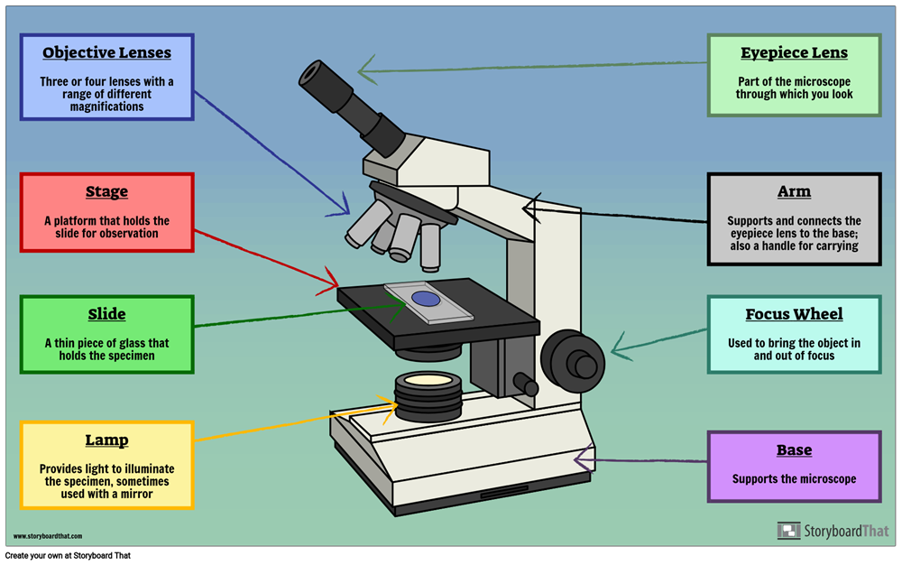
Parts of the Microscope Label and Definition Diagram | Quizlet Medium Power Objective. Provides magnification, usually about 10x; total magnification is 100. High Power Objective. Provides magnification, usually about 40x; total magnification is 400. Stage Clips. Grip slide in place for. viewing. Diaphragm. Controls amount of light entering the body tube.
Microscope diagram with labels and definitions
A physical wiring diagram for the human immune system | Nature Aug 03, 2022 · For imaging, a PerkinElmer Opera Phenix automated spinning-disk confocal microscope was used and each well of a 348-well plate was imaged at 20× magnification with 5 × 5 non-overlapping images ... Microscope Types (with labeled diagrams) and Functions Simple microscope labeled diagram Simple microscope functions It is used in industrial applications like: Watchmakers to assemble watches Cloth industry to count the number of threads or fibers in a cloth Jewelers to examine the finer parts of jewelry Miniature artists to examine and build their work Also used to inspect finer details on products PDF Definitions of the Parts of the Microscope - UAlberta microscope moves the stage up and down to bring the specimen into focus. The gearing mechanism of the adjustment produces a large vertical movement of the stage with only a partial revolution of the knob. Because of this, the coarse adjustment should only be used with low power (4X and 10X objectives) and never with the high power lenses (40X and
Microscope diagram with labels and definitions. Parts of a microscope with functions and labeled diagram - Microbe Notes Microscope Definition Microscopes are instruments that are used in science laboratories to visualize very minute objects such as cells, and microorganisms, giving a contrasting image that is magnified. Microscopes are made up of lenses for magnification, each with its own magnification powers. Electron Microscope-Definition, Principle, Types, Uses, Labeled Diagram Parts of an Electron Microscope The electron microscope is placed vertically and has the shape of a tall vacuum column. It consists of the following elements: 1. Electron gun A heated tungsten filament that produces electrons makes up the electron cannon. 2. Electromagnetic lenses The condenser lens directs the electron beam to the specimen. Microscopy- History, Classification, Terms, Diagram - The Biology Notes History of Microscope. In the 1 st Century AD, the Romans invented the glass and used them to magnify objects. In the early 14 th Century AD, eyeglasses were made by Italian spectacle makers. In 1590, two Dutch spectacle makers, Hans, and Zacharias Jansen created the first microscope. It was a simple tube with 2 lenses system and had 9X ... Science S1 Flashcards | Quizlet Study with Quizlet and memorize flashcards containing terms like In which order did the events forming our solar system occur? The solar nebula became hot and dense pulling in more gas.This flattened into a rotating disk.
Types of Microscopes: Definition, Working Principle, Diagram ... - BYJUS There are also microscope types that find application in metallurgy and studying three-dimensional samples. In this article, there are 5 such microscope types that are discussed along with their diagram, working principle and applications. These five types of microscopes are: Simple microscope. Compound microscope. Label Microscope Diagram Flashcards | Quizlet A small lens with low magnigying power Mirror (or light source) This directs light upwards onto the slide Revolving Nosepiece The rotating device that holds the objectives (lenses) Stage The platform on which a slide is placed. Stage Clips Metal clips that hold a slide securely onto the stage. full microscope diagram Sets with similar terms Microscope: Types of Microscope, Parts, Uses, Diagram - Embibe Microscope: Definition, Parts, Parameters, Types, & Uses A microscope is an instrument that produces enlarged images of small objects. It provides the observer an exceedingly close view of minute structures at a scale convenient for examination and analysis. The microscope magnifies microscopic objects that are not visible to the naked eyes. ISO/IEC Guide 99:2007(en), International vocabulary of ... The second edition of the International vocabulary of basic and general terms in metrology (VIM) was published in 1993. The need to cover measurements in chemistry and laboratory medicine for the first time, as well as to incorporate concepts such as those that relate to metrological traceability, measurement uncertainty, and nominal properties, led to this third edition.
Microscope, Microscope Parts, Labeled Diagram, and Functions Microscope, Microscope Parts, Labeled Diagram, and Functions What is Microscope? A microscope is a laboratory instrument used to examine objects that are too small to be seen by the naked eye. It is derived from Ancient Greek words and composed of mikrós, "small" and skopeîn,"to look" or "see". Compound Microscope Parts, Functions, and Labeled Diagram Compound Microscope Parts, Functions, and Labeled Diagram Parts of a Compound Microscope Each part of the compound microscope serves its own unique function, with each being important to the function of the scope as a whole. Compound Microscope Parts - Labeled Diagram and their Functions The eyepiece (or ocular lens) is the lens part at the top of a microscope that the viewer looks through. The standard eyepiece has a magnification of 10x. You may exchange with an optional eyepiece ranging from 5x - 30x. [In this figure] The structure inside an eyepiece. The current design of the eyepiece is no longer a single convex lens. E-Learning – AOAC India Bright Field Microscope, Dark Field Microscope, Phase Contrast Microscope, Fluorescence Microscope, Confocal microscopy, Scanning and Transmission Electron Microscope and applications: 17th June 2019, 2019 11.30-12.30 pm: Watch Video: 50 : Nuclear magnetic resonance (NMR) – Part 1 DR. CHANDRASHEKHAR MR. RAGHAV MAVINKURVE, BRUKER
Labelled Microscope with Functions Storyboard Szerint oliversmith
16 Parts of a Compound Microscope: Diagrams and Video Body of the Microscope In compound microscopes with two eye pieces there are prisms contained in the body that will also split the beam of light to enable you to view the image through both eye pieces. 2. Arm The arm of the microscope is another structural piece. The arm connects the base of the microscope to the head/body of the microscope.

PPT - Plant Cell Journal - Elodea PowerPoint Presentation - ID:1159196
A Study of the Microscope and its Functions With a Labeled Diagram ... A Study of the Microscope and its Functions With a Labeled Diagram To better understand the structure and function of a microscope, we need to take a look at the labeled microscope diagrams of the compound and electron microscope. These diagrams clearly explain the functioning of the microscopes along with their respective parts.

The Microscope: Create a Labelled Diagram | Teaching Resources
(PDF) Cambridge International AS and A Level Biology ... BIO1: Maintaining a Balance 1. Most organisms are active in a limited temperature range IDENTIFY THE ROLE OF ENZYMES IN METABOLISM, DESCRIBE THEIR CHEMICAL COMPOSITION AND USE A SIMPLE MODEL TO DESCRIBE THEIR SPECIFICITY ON SUBSTRATES
Plant Cell Anatomy - EnchantedLearning.com
Compound Microscope: Definition, Diagram, Parts, Uses, Working ... - BYJUS A compound microscope is defined as. A microscope with a high resolution and uses two sets of lenses providing a 2-dimensional image of the sample. The term compound refers to the usage of more than one lens in the microscope. Also, the compound microscope is one of the types of optical microscopes. The other type of optical microscope is a ...

All Saints Online: Diagram for Labelling: Microscope
Parts of Stereo Microscope (Dissecting microscope) - labeled diagram ... Labeled part diagram of a stereo microscope Major structural parts of a stereo microscope Optical components of a stereo microscope - definition and function Eyepieces Eyepiece tube Diopter adjustment ring Interpupillary Adjustment Objective Lenses Barlow lens Adjustment Knobs Light sources Stage plate Stage chips
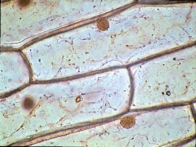
8.1 & 8.2 Math and Science: Onion Skin Lab
Laboratory procedures for diagnosis of anthrax, and isolation ... 1. Anthrax and the microbiology laboratory; operational safety. With some country-to-country variation in safety level definitions and requirements, recommendations for the manipulation of the causative agent of anthrax, Bacillus anthracis, generally are that BSL (biosafety level) 2 practices, containment equipment and facilities are appropriate for diagnostic tests, but BSL3 standards should ...

All Saints Online: Diagram for Labelling: Microscope
Microscope Parts & Functions - AmScope Microscope Terms. This is a glossary of commonly used microscopy terms. Abbe Condenser: A lens that is specially designed to mount under the stage and which typically moves in a vertical direction. An adjustable iris controls the diameter of the beam of light entering the lens system. Both by changing the size of this iris and by moving the ...
14 Best Images of Labeled Plant Cell Parts Worksheet - Prokaryotic Cell Coloring Page, Label ...
microscope | Types, Parts, History, Diagram, & Facts microscope, instrument that produces enlarged images of small objects, allowing the observer an exceedingly close view of minute structures at a scale convenient for examination and analysis. Although optical microscopes are the subject of this article, an image may also be enlarged by many other wave forms, including acoustic, X-ray, or electron beam, and be received by direct or digital ...

257 best images about Science Teaching ideas! on Pinterest | Science classroom, Plate tectonics ...
Label the microscope — Science Learning Hub In this interactive, you can label the different parts of a microscope. Use this with the Microscope parts activity to help students identify and label the main parts of a microscope and then describe their functions. Drag and drop the text labels onto the microscope diagram.

Minds Eye: August 2015
Parts of the Microscope with Labeling (also Free Printouts) Parts of the Microscope with Labeling (also Free Printouts) By Editorial Team March 7, 2022 A microscope is one of the invaluable tools in the laboratory setting. It is used to observe things that cannot be seen by the naked eye. Table of Contents 1. Eyepiece 2. Body tube/Head 3. Turret/Nose piece 4. Objective lenses 5. Knobs (fine and coarse) 6.

Cross Section Of A Plant Cell - slidesharetrick
PDF Parts of a Microscope Printables - Homeschool Creations Label the parts of the microscope. You can use the word bank below to fill in the blanks or cut and paste the words at the bottom. Microscope Created by Jolanthe @ HomeschoolCreations.net. Parts of a eyepiece arm stageclips nosepiece focusing knobs illuminator stage objective lenses

Scope Body Parts Microscope Parts & Accessories - Boli Optics Microscopes & Accessories
Compound Microscope- Definition, Labeled Diagram, Principle, Parts, Uses Compound Microscope- Definition, Labeled Diagram, Principle, Parts, Uses April 3, 2022 by Sagar Aryal What is a Compound Microscope? Working Principle of the Compound Microscope Magnification of compound microscope Parts of a Compound Microscope Objectives and Stage Clips Arm and Base Illuminator and Stage Nosepiece and Aperture

29 best Anatomy and Physiology images on Pinterest | Physiology, Anatomy and Anatomy reference
Label Microscope Diagram - EnchantedLearning.com low-power objective - a small lens with low magnifying power. mirror (or light source) - this directs light upwards onto the slide. revolving nosepiece - the rotating device that holds the objectives (lenses). stage - the platform on which a slide is placed. stage clips - metal clips that hold a slide securely onto the stage.

16 Parts of the Microscope ideas | microscope, microscopic, microscope parts
Microscope Parts and Functions First, the purpose of a microscope is to magnify a small object or to magnify the fine details of a larger object in order to examine minute specimens that cannot be seen by the naked eye. Here are the important compound microscope parts... Eyepiece: The lens the viewer looks through to see the specimen.

Honors Biology Midterm Flashcards | Easy Notecards
CODEX multiplexed tissue imaging with DNA-conjugated ... - Nature Jul 02, 2021 · A newly adapted version of CODEX uses an automated microfluidics system and conventional fluorescent microscope to iteratively hybridize, image and strip fluorescently labeled DNA probes that are ...

The Animal Cell; What is the composition and functionality? by Ben Lieberman | Animal cell ...
Microscope Parts, Function, & Labeled Diagram - slidingmotion Microscope parts labeled diagram gives us all the information about its parts and their position in the microscope. Microscope Parts Labeled Diagram The principle of the Microscope gives you an exact reason to use it. It works on the 3 principles. Magnification Resolving Power Numerical Aperture. Parts of Microscope Head Base Arm Eyepiece Lens
Post a Comment for "45 microscope diagram with labels and definitions"